.png)
.png)
A lesion is any area of tissue that looks or behaves differently from the surrounding healthy tissue. It can be visible on the skin, found inside an organ, or detected on an imaging test, and it can arise from injury, infection, inflammation, abnormal growth, or healing. Because the word is a broad medical term rather than a diagnosis, understanding context—where it is, what caused it, and how it acts—matters far more than the label itself. By the end of this guide, you’ll know how clinicians describe lesions, how they are evaluated, and what common treatment paths look like.
At its core, the word lesion simply means “an area of change.” That change might be a patch of irritated skin, a small cyst, a polyp in the colon, a spot on a lung scan, or scar tissue after an injury. Some lesions are temporary and harmless; others signal a condition that needs attention. Context clues—such as size, shape, borders, growth over time, symptoms, and risk factors—help providers decide whether a lesion can be watched or should be tested and treated.
Lesions form when normal cells or structures are disrupted. Triggers include trauma, infections, autoimmune reactions, clogged ducts, degenerative processes, and uncontrolled cell growth. After the initial trigger, the body’s response—swelling, repair, scarring, or overgrowth—shapes the lesion's appearance and behavior.
An acute injury can damage cells, leading to swelling, redness, and tenderness as the body heals. With repeated irritation, lingering inflammation can create thickened or scarred areas.
Bacteria, viruses, fungi, and parasites can create localized damage or fluid collections. The immune system’s effort to contain invaders may leave behind noticeable changes.
Sometimes cells grow too quickly or fail to die on schedule, forming masses or nodules. In other cases, tissue breaks down or loses function, leaving thin, weak, or discolored areas.
Lesions can appear almost anywhere, from the body’s surface to deep internal organs. Their impact depends on location: a small spot in a critical structure can cause more trouble than a larger one in a less sensitive area. Location also guides the best test to use and whether treatment is urgent or elective.
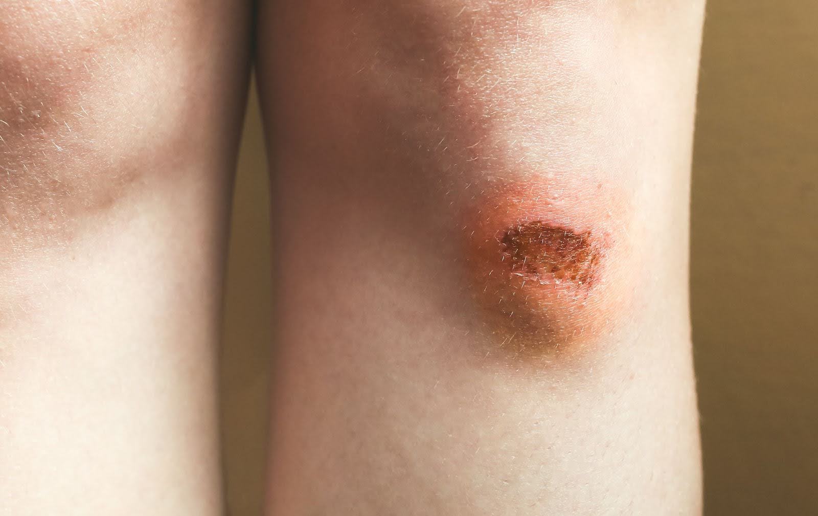
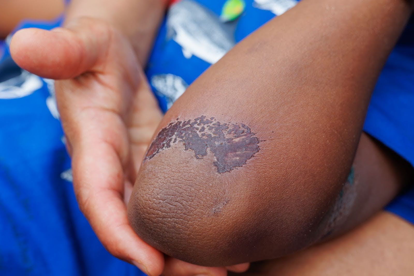
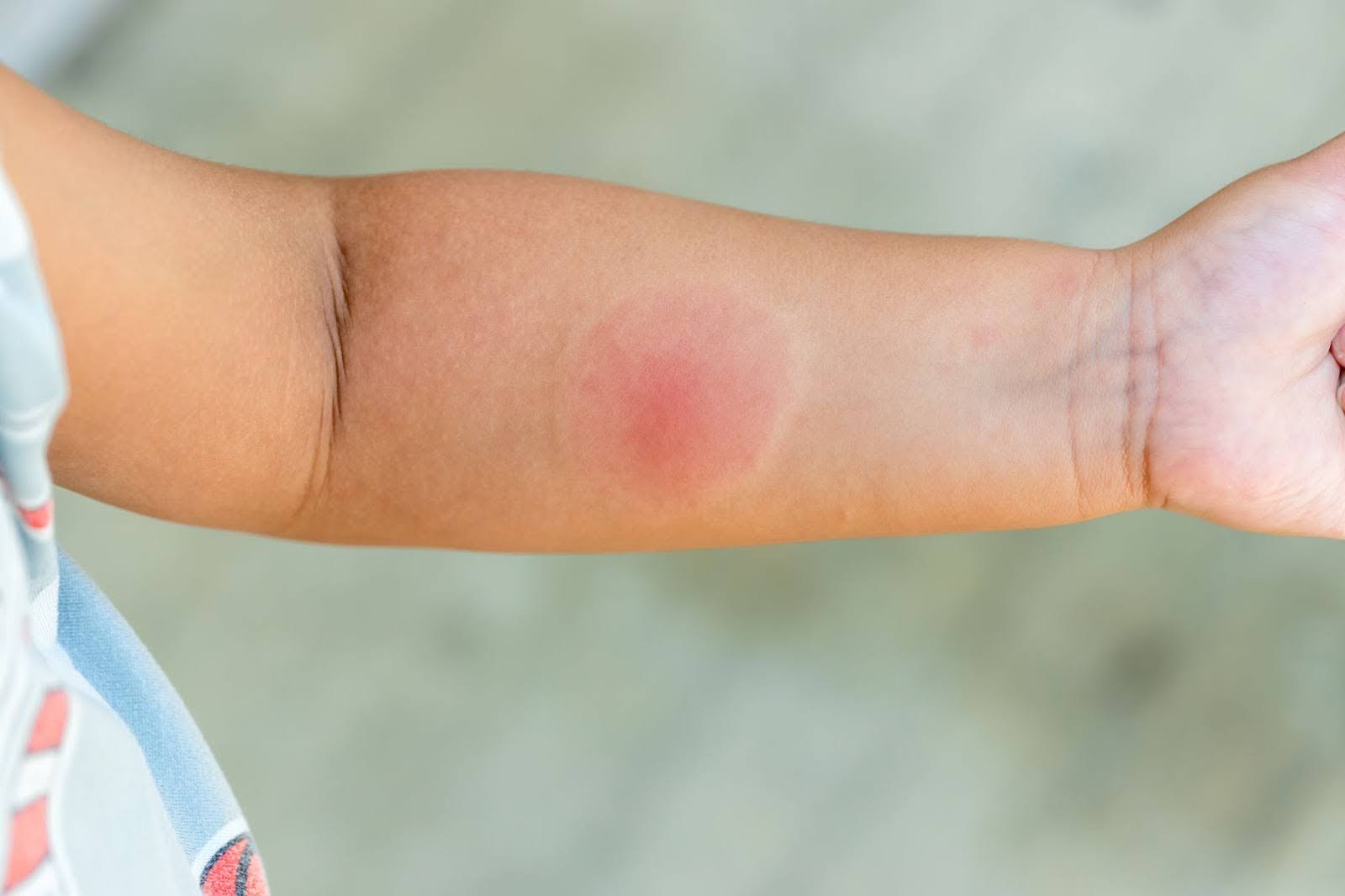
Spots, bumps, ulcers, and rashes on the skin or mouth are among the most visible lesions. These often come from irritation, allergies, infections, or sun exposure.
Lesions in the nervous system may affect movement, sensation, or cognition. Even tiny changes in key areas can create noticeable symptoms.
H3: Heart and Blood Vessels
Plaques, scars, or clots can disrupt blood flow and strain the heart. Vascular lesions may present with pain, swelling, or changes in skin color.
Spots seen on chest imaging might be scars, infections, or growths. The pattern, size, and evolution over time help determine their significance.
Polyps, ulcers, and inflamed patches can develop in the esophagus, stomach, or intestines. Some are incidental findings; others cause pain, bleeding, or changes in bowel habits.
Clinicians describe lesions using features that predict behavior. The words can sound technical, but they boil down to a few key ideas: what’s inside, how it looks, how it grows, and how long it has been present.
Solid lesions are made of tissue, while cystic ones contain fluid or semi-fluid material. This difference affects both testing and treatment choices.
Benign lesions tend to grow slowly and stay localized. Malignant ones invade nearby structures and can spread, which is why careful evaluation matters.
Acute lesions arise quickly and often relate to injury or infection. Chronic ones linger, wax and wane, or slowly progress over time.
It’s common to notice lesions on the skin because they are visible and easy to monitor. Acne nodules, cold sores, warts, eczema patches, moles, and scars are everyday examples, and most are not serious. Red flags to watch for include moles that change shape or color, sores that don’t heal, rapidly growing lumps, and rashes accompanied by fever or severe pain. Photographs taken weeks apart help track change and guide decisions about when to be seen.
Plenty of lesions live out of sight and show up only on scans or scopes. A polyp found during a colonoscopy, a thyroid nodule on ultrasound, a small spot on a liver MRI, or a white matter change on a brain MRI can all be lesions with very different implications. Many are incidental and harmless, discovered while looking for something else. Others guide preventive care, such as removing a colon polyp to reduce future cancer risk or following liver spots with periodic imaging.
Evaluation starts with a story and an exam, then proceeds to targeted tests. The goals are to define the lesion’s structure, rule in or out concerning causes, and match findings with the right level of intervention. A measured approach avoids both over-testing and missed diagnoses, balancing peace of mind with practicality.
Providers ask about timing, triggers, symptoms, and change. They examine size, borders, texture, warmth, and tenderness to narrow the list of possibilities.
Ultrasound, X-ray, CT, and MRI each reveal different details. Comparing current and prior images shows whether a lesion is stable, shrinking, or growing.
When answers remain uncertain, a sample may be taken for analysis. Blood work and cultures can also point toward infection, inflammation, or metabolic causes.
Treatment is tailored to what the lesion is doing, where it is, and the person’s overall health. In one case, observation is safest; in another, timely lesion removal prevents complications. Goals are consistent: relieve symptoms, prevent harm, and preserve function while minimizing side effects.
Some lesions are best watched with scheduled check-ins and repeat imaging. This avoids unnecessary procedures when the risk of change is low.
Topicals, oral medicines, injections, or image-guided procedures can shrink, calm, or drain specific lesions. These options often shorten recovery time.
When a lesion threatens function or shows high-risk features, removal or repair may be recommended. Pathology review confirms the exact diagnosis afterward.
Risk depends on many variables: age, genetics, environment, lifestyle, and existing conditions. While not every lesion is preventable, lowering exposure to known risks and keeping routine care up to date reduces the odds of serious problems. Prevention also means catching changes early so that treatment stays simple.
Sun protection, tobacco avoidance, safer work practices, and limiting harmful chemicals lower the risk. Nutrition, physical activity, and sleep support tissue repair.
Vaccines prevent infectious diseases caused by certain viruses. Age-appropriate screening tests catch precancerous or early lesions when treatment is most effective.
Managing diabetes, blood pressure, and autoimmune conditions reduces inflammation and scarring. Regular follow-up keeps minor issues from becoming bigger ones.
Most lesions are not emergencies, but a few features deserve quick attention. Rapid growth, severe pain, sudden bleeding, signs of infection, or symptoms that affect breathing, vision, strength, or speech warrant urgent evaluation. Trust your instincts—if something feels new and worrisome, a timely assessment is worthwhile.
A spot that expands quickly or changes hue can signal active disease. Early evaluation clarifies whether action is needed.
Pain out of proportion to the apparent cause or bleeding without an evident cause suggests deeper issues. These features are valuable clues for targeted testing.
Numbness, weakness, speech trouble, or chest pain can reflect lesions in critical organs. Immediate care helps protect function.
Many people live well with stable lesions that never cause trouble. The key is a simple plan: know what kind it is, understand what to watch for, and keep recommended appointments. Tracking symptoms and bringing questions to visits makes care collaborative, ensuring decisions match goals and values. If circumstances change—new medications, pregnancy, or upcoming travel—ask whether the monitoring plan should adapt.
Misconceptions can create unnecessary anxiety or false reassurance. Clearing them up helps you make practical, informed choices about testing and treatment, and it keeps focus on proven steps that protect health over the long term.
Most lumps and spots have benign causes. Careful evaluation sorts the harmless from the risky without assuming the worst.
Scar tissue is a standard repair product and fits the definition of a lesion. Its presence tells a story about healing, not just harm.
Photos and search results can mislead. A brief exam or test often answers questions more quickly and accurately.
Age changes both causes and responses to treatment. Children may develop lesions tied to growth, infections, or genetic patterns, while older adults more often face wear-and-tear changes, vascular issues, or medication effects. Plans are personalized to minimize risk and maintain quality of life at every stage.
In younger people, many lesions resolve as the immune system matures. When testing is needed, the least invasive options are preferred.
Thin skin, reduced reserve, and multiple medicines can complicate healing. Gentle treatments and close follow-up improve outcomes.
Medical notes use descriptive terms that may sound intimidating but are simply shorthand. Words like “hyperpigmented,” “ulcerated,” “calcified,” “indeterminate,” or “well-circumscribed” are neutral descriptors that guide next steps. When you hear them, ask what they mean for you—does this suggest observation, medication, a procedure, or nothing at all right now. Plain-language translations are part of reasonable care, and your team should welcome those questions.
The label lesion is a starting point, not a verdict. What matters next is understanding cause, location, behavior, and personal risk so you can choose the least invasive, most effective path. Most lesions are manageable, and many are harmless; the few that are not benefit from early attention. Partnering with your provider, keeping routine care up to date, and speaking up when something changes are the simplest ways to stay ahead.
Visit our Naples Laser & Skin Aesthetics blog to learn more about lesions and how to treat them.
%402x.svg)